


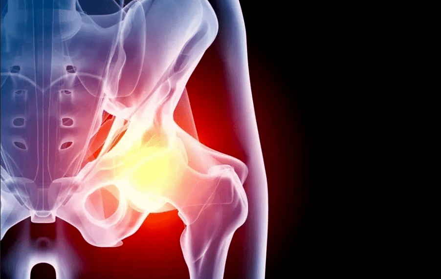
Arthrosis of the hip joint (coxarthrosis) is a chronic pathology, accompanied by gradual destruction of the cartilage tissue in the affected area, followed by the involvement of adjacent structures in the process. The disease requires long-term treatment and, in severe cases, the only way to regain mobility is joint replacement.
General information
Lumbarthrosis belongs to the group of degenerative diseases. It starts gradually with microscopic changes in the cartilage structure. Increased stress, inflammatory diseases, blood supply disorders lead to structural transformations and thinning of the cartilage tissue and, in turn, deform the contours of the joint region. As a result, the load distribution on the bone contact surfaces changes and the zones with maximum pressure begin to wear out more quickly. This triggers a whole cascade of pathological reactions:
- the appearance of micro-cracks and areas of compaction in the cartilaginous tissue;
- decreased smoothness of joint surfaces;
- cartilage overgrowth at the site of thinning and replacement by bone tissue;
- the appearance of osteophytes (bone growths) along the edges of the joint site;
- thickening and decreased elasticity of the joint capsule;
- hardening and reduced strength of ligaments;
- changes in synovial fluid composition (natural lubrication within the joint);
- joint space narrowing;
- fusion of all elements of the joint (ankylosis).
Without treatment, coxarthrosis inevitably becomes the cause of immobility and disability.
Causes
Depending on the causes of disease development, a distinction is made between primary and secondary coxarthrosis. In the first case, it occurs by itself, for example, in the context of an inherited predisposition; in the second, it is caused by other illnesses or injuries. In most cases, the cartilage tissue degeneration process occurs due to a combination of several factors. The reason could be:
- Congenital dislocation of the hip;
- flat feet, scoliosis and other orthopedic problems;
- Legg-Calve-Perthes Disease;
- joint inflammation (arthritis), regardless of origin;
- hip joint injuries and microtraumas in the context of overweight, professional sports, etc. ;
- hip joint dysplasia;
- metabolic disorders;
- endocrine disorders (especially diabetes mellitus);
- disturbances of blood supply to the lower extremities;
- frequent stress;
- heredity (coxarthrosis in parents or other close relatives significantly increases the risk of developing it in a child);
- congenital pathologies and autoimmune diseases of the connective tissue (joint hypermobility, rheumatoid arthritis, systemic lupus erythematosus, etc. );
- submitted to joint operations.
Age is an important predisposing factor. According to statistics, after 45 years, the probability of developing coxarthrosis increases significantly.
Symptoms
The main symptoms of hip joint coxarthrosis do not depend on the cause of development. Most patients observe:
- restriction of movement: one of the first symptoms due to thinning of the cartilage layer and increased friction of the articular surfaces of bones; in the future, the appearance of cartilaginous growths aggravates the problem even more;
- pain: friction of bones deprived of a cartilaginous layer against each other, gradual involvement of all elements of the joint in the degenerative process, decreased blood supply to the tissues causing sensations of pain that increase as the disease progresses; the pain is acute and often worsens later in the day;
- muscle spasm, which leads to increased symptoms of pain and limited joint range of motion;
- decreased leg length: this symptom appears in the later stages of the disease due to the narrowing of the joint space and the gradual grinding of the heads of bones due to constant friction; the difference between the legs can be up to 2 cm;
- lameness: associated with severe pain and limited movement, as well as leg shortening; it is an unfavorable sign that indicates serious damage to the joint apparatus.
Stages
In the development process, coxarthrosis goes through several stages, which depend on the degree of tissue damage.
- 1 degree. At this point, the patient notices mild pain in the joint that appears after intense or prolonged physical activity and quickly subsides after rest. As a rule, discomfort occurs precisely in the region of the hip joint, but in some cases it extends to the hip or knee. Gait does not change, leg movements are fully preserved. On radiography, specific changes are observed: subchondral sclerosis.
- 2nd degree. The pain becomes stronger, appears after exertion, spreading throughout the thigh and groin. After the effort, a slight lameness may appear. Difficulties in abducting the leg are encountered. The radiograph shows a significant decrease in the distance between the bones (by 50% or more), deformation of the femoral head and pronounced bone growths.
- 3 degrees. The pain becomes permanent, walking without a cane becomes impossible. When moving, the patient visibly leans to the painful side, which further increases the load on the joint. Range of motion is reduced, leg and buttock muscles atrophy. There is a shortening of the affected limb. The radiograph reveals a significant deformity of the joint, changes in the contour of the femoral head, and a large number of osteophytes.
- 4 degrees. The pain gets stronger and doesn't stop for a minute, the patient loses the ability to move independently. The radiograph shows complete destruction of the articular cartilage, as well as signs of bone fusion with each other (ankylosis). Dealing with the disease at this stage is only possible through surgery.
Diagnosis
An orthopedic traumatologist is responsible for identifying symptoms and selecting treatment. To diagnose and determine the extent of the disease, he uses:
- research: listening to patients' complaints, identifying risk factors (trauma, disease, heredity, etc. );
- exam: evaluation of limb mobility, determination of areas of greatest pain;
- X-ray: X-ray makes it possible to assess the state of bones and cartilage, the size of the joint space, the presence and location of bone growths; to see the necessary details in more detail, the study is complemented by CT (computed tomography);
- laboratory diagnosis: a general blood test allows you to identify signs of an inflammatory, biochemical process - look at some risk factors, eg uric acid level;
- MRI (magnetic resonance): allows you to assess the condition not only of bones and cartilage, but also of soft tissues: bones, ligaments, muscles, joint capsule, etc. ;
- puncture of the joint.
If it is necessary to perform differential diagnoses with other diseases, as well as to evaluate concomitant pathologies, complementary exams, instrumental exams and consultations with restricted specialists are prescribed.
Coxarthrosis Treatment
Treatment of hip joint coxarthrosis depends on the stage and severity of symptoms. The pathology requires an integrated approach using several methods:
- drug treatment;
- non-drug treatment (physiotherapy, exercise therapy);
- surgery;
- lifestyle and diet correction.
drug treatment
The drugs prescribed for arthrosis of the hip joint aim to:
- removal of pain syndrome;
- restoring or at least slowing the destruction of cartilage tissue;
- improve blood supply and nutrition to the affected area;
- treatment of concomitant pathologies.
Analgesics are used in the form of tablets, intramuscular and intra-articular injections and topical agents: creams, ointments, patches. In the early stages of disease development, nonsteroidal anti-inflammatory drugs are sufficient for most patients. In severe pain syndrome, hormonal agents are used. The introduction of analgesics directly into the joint capsule has a good effect.
If the course of the disease is accompanied by muscle spasm, muscle relaxants are used. They are used in combination with other pain relievers.
Taking analgesics should be limited in time and dose, so as not to cause further damage to cartilage tissue and other side effects (in particular, the development of gastritis and stomach ulcers).
Chondroprotective drugs are medicines that help to restore cartilage tissue. They are effective only with regular long-term use, which is combined with other treatment methods, lifestyle, and dietary adjustments. Medicines to improve blood microcirculation help to increase its effect. For a similar purpose, warming ointments are prescribed. Only one physician is involved in dosage and regimen selection.
drug-free treatment
This category includes various physical therapy techniques and manuals as well as physical therapy exercises. They help to improve microcirculation and restore movement to the damaged joint. Depending on the situation, the doctor prescribes:
- shock wave therapy;
- magnetotherapy;
- electromyostimulation;
- various types of electrophoresis and phonophoresis (accompanied by the administration of anesthetic drugs);
- mechanotherapy;
- massage and exercise therapy.
Surgery
If the disease has reached stage 3-4 of development, medications and physical therapy will only alleviate the patient's condition, but will not restore the patient's ability to move fully. In this case, arthroplasty is indicated, that is, complete or partial replacement of the damaged joint with a titanium prosthesis.
If there are indications, a lighter version of the intervention is performed: sanding of bone contact areas and covering with special smooth implants that facilitate sliding.
Prevention
Lifestyle can significantly reduce the risk of developing coxarthrosis, as well as the rate of its progression. It is important to strictly comply with the rules:
- lead an active lifestyle: swimming in the pool, walking, cycling - amateur-level physical activity without racing to records helps improve blood supply and inhibits joint degeneration processes;
- normalize body weight to reduce load on legs;
- eliminate injuries, hypothermia and occupational risk factors (vibration, weight lifting, standing work);
- timely treat all diseases, including those not directly related to the musculoskeletal system;
- correct posture disorders in time, wear comfortable shoes.
Diet
With the help of nutritional correction, the patient can not only reduce body weight, but also reduce inflammatory reactions, tissue salt deposits and metabolic disturbances. It is recommended that you follow a balanced menu with sufficient but not excessive amounts of carbohydrates, proteins and fats, as well as vitamins and minerals. Special attention should be paid to unsaturated fats (olive oil and flaxseed), omega-3 acids (found in excess in fish), collagen (gelled meat, peas). It is recommended to minimize fast carbohydrates, alcohol, strong coffee, products with artificial flavors, preservatives and flavor enhancers.
Consequences and complications
Lumbarthrosis is one of the common causes of disability in elderly people. Without proper treatment, the pathology inevitably leads to total disability, especially in bilateral injuries. The pain and difficulty in walking do not allow you to work and take care of yourself, so it is important to get treatment at the right time.